
X-ray, CT and MRI rooms for clinics, independent hospitals and imaging providers
Clinical Visions designs and builds diagnostic imaging rooms for clinics, independent hospitals and imaging providers: X-ray and CT rooms shielded under IRR17 to the radiation protection adviser’s specification, MRI rooms planned around the MHRA’s access zones, and the controlled areas, cooling and power every modality depends on, phased around a working department.
- Shielding built to the radiation protection adviser’s specification
- MRI planned around the controlled access area from the first drawing
- Phased so the rest of the department keeps scanning
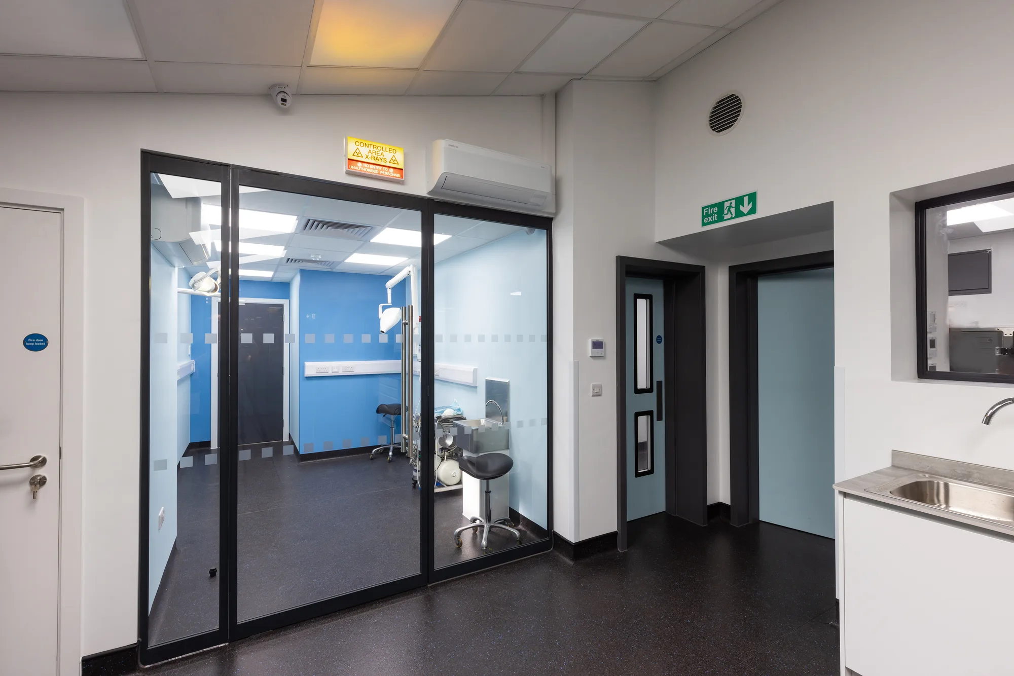
Shielding that is right before the plaster goes on
Once an imaging room is finished, its shielding cannot be seen. Every lapped joint, every service penetration and every door frame is hidden behind the finishes, so it has to be right, and recorded, at the point it is installed.
The record of what was installed, and where, is handed over with the room for the adviser’s survey.
- The radiation protection adviser’s specification fixed before anything is drawn
- Shielding continuous at joints, frames, penetrations and service boxes
- Each wall and ceiling inspected and photographed before it is closed
- MRI shielding and access zones coordinated with the scanner supplier
- Controlled areas marked out and signed as the regulations require
Every modality is a different building problem
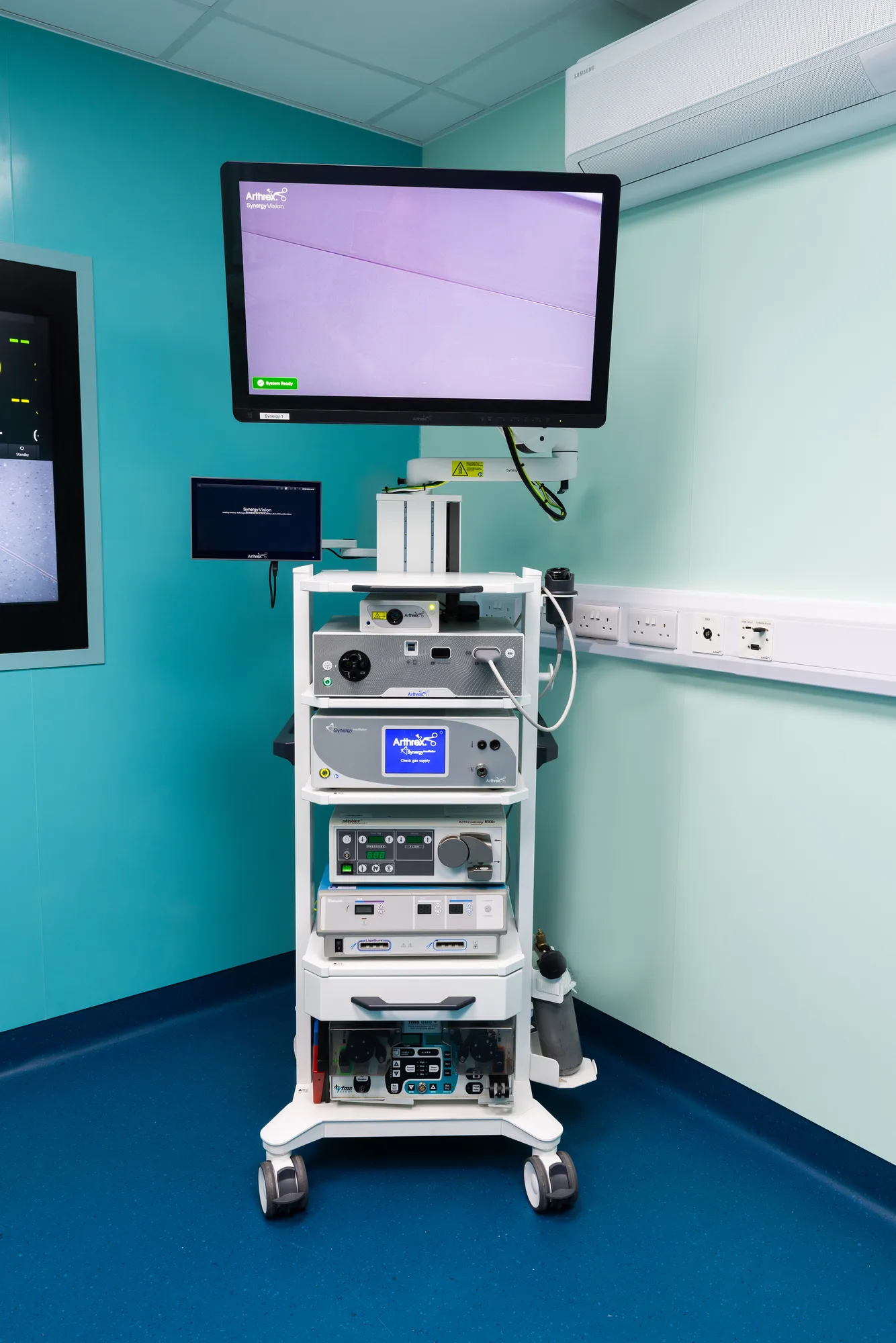
An X-ray room, a CT room and an MRI suite share a department and almost nothing else.
X-ray, fluoroscopy and CT use ionising radiation, so the room is a shielded box: shielding in the walls, the doors, the windows and sometimes the floor and ceiling, continuous at every joint and penetration, and a controlled area marked out around it. MRI uses a magnetic field and radio waves and needs none of that. It needs a radio-frequency shielded enclosure instead, a quench route for the magnet, and zones that keep anyone carrying metal well away from the scanner. Ultrasound needs neither, only privacy and the right light.
What every modality shares is the equipment. The scanner decides the room size, the floor loading, the cooling and the route the equipment takes into the building, so the supplier’s site requirements are fixed before anything else is drawn. A room designed first and fitted with a scanner afterwards is a room rebuilt.
The regulations that shape an imaging room
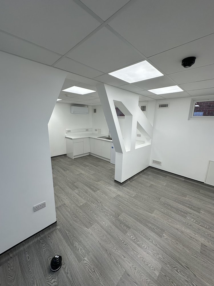
Two regulations cover ionising radiation, and in a medical building both apply.
IRR17, the Ionising Radiations Regulations 2017, protects staff and the public. It is the regulation that shapes the building: the employer’s risk assessment, the radiation protection adviser who specifies the shielding, and the controlled areas that are designated, marked and signed. IR(ME)R, the Ionising Radiation (Medical Exposure) Regulations 2017, protects the patient being examined. It applies to medical imaging and does not apply to veterinary practice, which is the single most important difference between a medical imaging room and one in a veterinary hospital.
MRI sits outside both. The MHRA safety guidelines for MRI equipment in clinical use set out the MR Controlled Access Area and the MR Environment, and the suite is planned around them. An imaging service in England is usually a regulated activity, so CQC registration and the regulation on premises and equipment apply, and NHS England’s HBN 6 on facilities for diagnostic imaging is the planning reference for how a department is laid out.
Where imaging sits in a clinic project
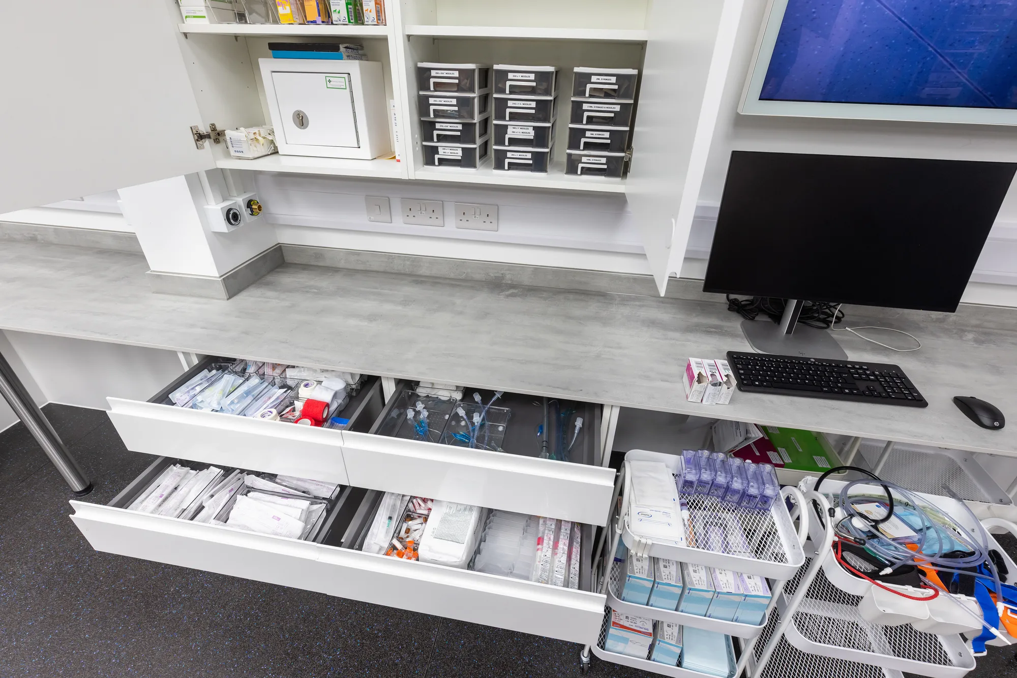
Sometimes it is the whole project. More often it is one room added to a clinic that is already open.
A new X-ray or CT room inside a working medical clinic is built the same way as any phased clinical work: sealed off, noisy work timed around clinic hours, the delivery route planned and protected. The shielding detail that decides whether the room passes its survey is the same as in any lead-lined imaging room: continuous at every joint, frame and penetration. The power, cooling and data each scanner needs are designed with the mechanical and electrical package, and a new department in a building that has never had one usually starts as design and build, so the structure, the plant and the shielding are designed by the same team that builds them.
Standards governing this sector
The requirements that shape the building, each quoted from the regulator that will inspect it and linked to the source.
| Code | Standard | What it requires | Source |
|---|---|---|---|
| IRR17 reg 8 | Ionising Radiations Regulations 2017, regulation 8, radiation risk assessments | An employer, before commencing a new activity involving work with ionising radiation in respect of which no risk assessment has been made by that employer, must make a suitable and sufficient assessment of the risk to any employee and other person for the purpose of identifying the measures the employer needs to take to restrict the exposure of that employee or other person to ionising radiation. | legislation.gov.uk |
| IRR17 reg 14 | Ionising Radiations Regulations 2017, regulation 14, radiation protection adviser | Every employer engaged in work with ionising radiation must consult such suitable radiation protection advisers as are necessary for the purpose of advising the employer on the observance of these Regulations, must appoint that adviser in writing, and must include in that appointment the scope of the advice which the radiation protection adviser is required to give. | legislation.gov.uk |
| IRR17 reg 17 | Ionising Radiations Regulations 2017, regulation 17, designation of controlled or supervised areas | The employer must designate as a controlled area any area where a person must follow special procedures to restrict significant exposure, or where a person is likely to receive an effective dose greater than 6 mSv a year. | legislation.gov.uk |
| IRR17 reg 19 | Ionising Radiations Regulations 2017, regulation 19, additional requirements for designated areas | A designated area must be described in the local rules and marked with suitable and sufficient signs warning of the designation and the nature of the radiation source, and a controlled area must be physically demarcated or, where that is not reasonably practicable, delineated by other suitable means. | legislation.gov.uk |
| IR(ME)R reg 3 | Ionising Radiation (Medical Exposure) Regulations 2017, regulation 3, application | The regulations apply to exposure to ionising radiation of patients as part of their own medical diagnosis or treatment, of individuals in health screening programmes, of patients or other persons voluntarily taking part in medical or biomedical research, of carers and comforters, of asymptomatic individuals, and of individuals undergoing a non-medical imaging exposure. | legislation.gov.uk |
| IR(ME)R reg 15 | Ionising Radiation (Medical Exposure) Regulations 2017, regulation 15, equipment general duties of the employer | The employer must carry out adequate testing of any equipment before it is first used for a medical exposure, performance testing at regular intervals, and performance testing following a maintenance procedure which is capable of affecting the equipment's performance. | legislation.gov.uk |
| MHRA MRI guidelines, 4.2.1 | Medicines and Healthcare products Regulatory Agency, Safety Guidelines for Magnetic Resonance Imaging Equipment in Clinical Use (fifth edition, 2026), MR Controlled Access Area | Each MRI unit should define an MR Controlled Access Area large enough to contain the MR Environment, with access restricted, suitable signage at every entrance, doors kept closed except when in use, entrances kept to a minimum, and unrestricted access given only to MR Authorised Persons. | GOV.UK |
| CQC registration | Care Quality Commission, quick guide to regulated activities | A provider carrying on a regulated activity such as treatment of disease, disorder or injury, surgical procedures or diagnostic and screening procedures must be registered with the Care Quality Commission, and a provider carrying on a regulated activity without being registered may be prosecuted and have to pay a fine. | CQC |
Who we build imaging rooms for
From a single X-ray room in a clinic to a multi-modality diagnostic department.
-
Diagnostic and imaging providers
-
Independent hospitals adding imaging
-
Community diagnostic centres
-
Private clinics and consulting suites
-
Sports medicine and orthopaedic clinics
-
Day-case and minor procedures units
How an imaging project runs
From the equipment outwards. The scanner and the adviser’s specification decide the room, so they are fixed first.
-
Agree the modalities and the equipment
Which scanners, from which supplier, in which rooms. The supplier’s site requirements set the room size, the structure, the cooling and the delivery route.
-
Fix the shielding and the zones
The radiation protection adviser specifies the shielding for X-ray and CT rooms. For MRI, the controlled access area and the MR Environment are drawn with the supplier.
-
Check the building can take it
Floor loading, structure, delivery route, plant space and the quench route for MRI, all checked on site before the design is fixed.
-
Build the shielding before the finishes
Shielding installed, inspected and recorded wall by wall, services sealed through it, then the room finished, in phases around the working department.
-
Hand over for the survey
The installed shielding record, test certificates and drawings as built handed over with the room, ready for the adviser’s survey and the equipment installation.
Figure
Where an X-ray or CT room has to stay continuous
A plan through a typical shielded imaging room, drawn to show where the shielding stops being a flat sheet and has to be lapped, backed or framed. The amount of shielding is the radiation protection adviser's calculation; the drawing shows where it must not break.
- 1 Shielded wall lining Lead-lined board or lead-backed sheet on a stud frame, built to the performance the adviser specifies for that particular wall.
- 2 Lapped corner and board joints Lead strip lapped behind every joint, stud line and internal corner, so the envelope is continuous rather than a set of shielded panels.
- 3 Shielded doorset Lead-lined leaf and frame with a lapped threshold, on hinges and a closer rated for the finished leaf weight.
- 4 Lead glass viewing panel Glazed into a lead-lined frame and positioned for the operator, with the frame lapped to the wall lining behind it.
- 5 Shielded control position Sited behind shielding with an unobstructed view of both the patient and the entrance to the room.
- 6 Backed service penetration Lead backing lapped behind every socket box, switch, data outlet and duct that passes through a shielded surface.
What we build in an imaging department
Each modality brings a different building problem, and a department brings all of them together. These are the rooms and the services we design and deliver.
X-ray and fluoroscopy rooms
General radiography and fluoroscopy rooms shielded to the adviser’s specification, with a protected control area, the controlled area marked out and warning signals at the doors.
CT rooms
Shielded scan rooms with the floor, the structure and the delivery route checked for the scanner, a control room with a view of the patient, and the cooling the equipment needs.
MRI suites
The radio-frequency shielded enclosure, quench route and access zones coordinated with the scanner supplier, so the MR Environment is contained inside the controlled access area.
Ultrasound, mammography and DEXA
The smaller rooms that complete a diagnostic service, each with the privacy, lighting and shielding, where it is needed, that its modality calls for.
Patient flow and support rooms
Waiting, changing, preparation and recovery arranged so a patient moves through the department in one direction, with reporting rooms kept away from the patient route.
Power, cooling and data
Supplies sized for the equipment, chilled water or dedicated cooling for scanners and equipment rooms, and data for image transfer, installed under our own F gas company certification.
Frequently Asked Questions
Does IR(ME)R apply to a medical imaging room?
Yes. IR(ME)R applies to the exposure of patients as part of their own medical diagnosis or treatment, so it governs every X-ray, fluoroscopy and CT examination a medical imaging room carries out. It sits alongside IRR17, which protects staff and the public and is the regulation that shapes the room. Veterinary imaging is different: IR(ME)R does not apply to veterinary practice, which sits under IRR17 alone.
Who decides how much shielding an X-ray or CT room needs?
The radiation protection adviser. The employer has to consult a suitable adviser under IRR17, and the adviser specifies the shielding from the equipment, the workload and the use of the rooms around it. We build to that specification, make it continuous at every joint and penetration, and record what was installed, so the adviser can survey the finished room against it.
Does an MRI room need lead shielding?
No. MRI does not use ionising radiation, so IRR17 shielding does not apply. An MRI room needs a radio-frequency shielded enclosure, sometimes magnetic shielding where the field would otherwise reach beyond the room, a quench route for the magnet, and the controlled access area described in the MHRA safety guidelines. All of it is coordinated with the scanner supplier before the room is designed.
Does a diagnostic imaging service need to register with the CQC?
Usually, in England. Diagnostic and screening procedures is a regulated activity, and a provider carrying it on must be registered with the Care Quality Commission. The premises then have to meet the regulation on premises and equipment, which is why the rooms are built to be clean, secure and suitable for their purpose. Check the CQC guidance for the exemptions that apply to your service.
Can an imaging room be built inside a working clinic?
Yes. The room is sealed off from the working clinic, noisy and dusty work is timed around clinic hours, and the scanner delivery route is planned and protected in advance, because it often runs through areas that stay in use. The rest of the department keeps scanning while the new room is built, shielded and handed over for the adviser’s survey.
What happens to the shielding records at handover?
They are handed over with the room. Each shielded wall, floor and ceiling is inspected and photographed before it is closed, and the record shows what was installed and where, alongside the drawings as built. That is the evidence the radiation protection adviser surveys the finished room against, and the reference for anyone who later drills, fixes or modifies it.
Get in Touch
Whether you're planning a complete refurbishment or a small specialist build, we’re here to help you create a safe, efficient, and modern clinical environment.